
Significant association of DNA variants with self-reported ME/CFS

Guest blog by Professor Chris Ponting and colleagues.
Summary
A new analysis using data from UK Biobank indicates that one version of a particular gene increases the risk of ME/CFS in women. The gene codes for a transporter protein in the mitochondrial membrane and plays a critical role in the urea cycle, which is important for removing ammonia from the body. Reduced levels of the transporter protein, which are expected for the gene variant associated with ME/CFS, are likely to impair mitochondrial function. If replicated later, this finding would provide the first evidence that a person’s risk for ME/CFS is caused by changes to mitochondrial function.
Background
On June 11 2018 we posted a blog describing an analysis of the UK Biobank’s data, drawn from half-a-million individuals from around the UK. The data implied that there is a genetic contribution to an individual’s risk of ME/CFS but it did not provide strong evidence that change in any one section of DNA explained this risk.
This analysis is called a genome-wide association study, or GWAS for short. When there is an association between a biological factor, such as cytokine changes, and an illness, it is not normally possible to say whether the factor is a cause of the illness or simply a consequence of being ill. But because the genetic variation comes first it must contribute to a cause rather than being an effect of the illness.

A GWAS asks whether the frequency of a DNA letter difference predicts whether a person has, or has not, a disease;
This prediction is not perfect and almost always reflects only a slight preference for people with this DNA difference to part of the disease cohort;
Importantly, GWAS predictions indicate genetic cause. This is because inherited disease (except cancer) does not predictably cause DNA mutation.
We are blogging again because a new analysis has revealed a promising finding.
This new analysis is again of the UK Biobank data and has been posted by Ben Neale’s lab. After discarding data that they considered lower quality, they were left with data from 194,174 females and 167,020 males and kindly made their results freely available to all.
Female-only GWAS
Considering males and females together they identified no specific region of the human genome whose DNA variants were significantly associated with self-reported CFS/ME. (One variant, rs148723539, is possibly indicated [p = 2.3×10⁻⁹] but this is not supported by adjacent variants.)
However, the female-only analysis revealed a single region, on chromosome 13. Ten DNA variants (single nucleotide variants, SNVs) were significantly associated [SNVs with minor allele frequency <0.001 or that were “low confidence” were filtered out] using a probability threshold of p < 5×10⁻⁸.
These 10 SNVs are inherited down the generations together (they are in “linkage disequilibrium” [LD]) and so this looks like just one association, rather than ten different ones. The 10 SNVs all lie in a 51,000 base region that surrounds the SLC25A15 gene (Figure 1 below).

Their conclusion is that DNA variation in this part of the genome slightly changes a woman’s risk of having a ME/CFS diagnosis. This must mean that one or more DNA differences in this part of the genome cause this risk change. But because all 10 differences are inherited together, it is not clear which one or ones are causing the increase in disease risk. Pinning down the causal DNA changes will require detailed experimental research.
We can talk about a DNA letter representing increased risk (this is the “risk allele”). For example, the 41,353,297th DNA letter on chromosome 13 is either G or A (the site is given the code rs7337312). You have two copies of chromosome 13, so also two versions of this DNA letter: you can have G twice (“GG”), A twice (“AA”) or G and A (“GA”). People across the world vary in the frequency of G or A but in the UK Biobank population it turns out that the frequency of G is about the same as that of A (~50%).
The Ben Neale lab result implies that having a DNA letter G at this position on chromosome 13 predicts a slight increase in risk of having a ME/CFS diagnosis; having two such letters G increases the risk further over having just one G. The frequency of G of ~50% means that roughly three-quarters of these UK Biobank females have at least one risk G.
Another of the Neale results is that women that have G at this position tend to have very slightly lower lymphocyte count (rs7337312; p = 4.6×10⁻⁷; other variants in LD have p < 5×10⁻⁸). Lymphocytes are white blood cells, mostly B cells and T cells. They also show that G at this position significantly and slightly increases two biomechanical properties of the cornea (corneal hysteresis and corneal resistance factor).

Ornithine Transporter type 1
Figure 1 shows that the GWAS genetic associations are for DNA differences that lie in-or-around a particular gene called SLC25A15. There is another gene in this Figure which is a pseudogene (TPTE2P5) which does not make protein and is less likely to alter physiological function when mutated. This does not necessarily immediately implicate SLC25A15 as the gene through which this genetic effect is transacted.
However, other data indicates that people that have G at this position tend to produce slightly lower amounts of SLC25A15 RNA (in aorta, colon, hippocampus, transformed lymphocytes and other samples, but not in whole blood, liver, muscle, cerebellum etc; Figure 2). So SLC25A15 is an excellent candidate for the gene whose activity alters between GG, GA and AA individuals thereby changing ME/CFS disease risk.
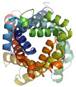
So what do we know about SLC25A15? Interestingly, it encodes a protein called Ornithine Transporter type 1 (ORNT1). This transports ornithine (as well as lysine and arginine) across the inner membrane of mitochondria to the mitochondrial matrix. Ornithine is an amino acid (but not incorporated into proteins) that plays a role in the urea cycle. This cycle plays an essential part in removing ammonia from the body (see point iv below).
This analysis predicts that if you have a letter G at this position then (Figure 2):
(i) if you are female, then you have a greater risk of ME/CFS;
(ii) many of your cells (for example in the heart or hippocampus, but not in muscle or liver) would tend to produce less ORNT1 RNA and less ORNT1 protein; and,
(iii) if so, then ornithine would build up in these cells and mitochondrial function overall would be impaired; and,
(iv) ammonia would accumulate in the blood.
Some of these predictions are drawn from what is known about individuals whose SLC25A15 genes are both defective, causing ornithine translocase deficiency (also known as HHH syndrome). This is a severe disease often leading to coma due to hyperammonemia, learning difficulties and also lethargy. A good review of HHH syndrome can be found here.
So do these predictions agree with what others have found when comparing people with ME/CFS with control individuals? In large part yes, but not always:
(a) Yamano et al. (2016) showed that there is an increased ornithine/citrulline ratio in ME/CFS individuals
(b) Naviaux et al. PNAS (2016) found that ornithine is moderately high for males and for females
(c) Nevertheless, Armstrong et al. (2012) found the opposite, a significant reduction of ornithine (P<0.05) in the blood of CFS samples.
What are this study’s limitations?
- Only one genomic region was identified, so a much larger study is required with about 20,000 ME/CFS cases to find more.
- This association would need replication in an independent study.
- We do not know whether males with ME/CFS show this genetic association. Males in UK Biobank with self-reported ME/CFS are 2.4-fold fewer than females, so a male-only GWAS is substantially less well powered to find true results.
- Even if true, then this association would alter risk for ME/CFS by only a small amount and certainly not for most people.
- The urea cycle occurs mostly in mitochondria in liver cells, yet SLC25A15 expression is found not to be different between GG, GA and AA in this organ. SLC25A15 is expressed also at moderate levels in pancreas and small intestine, yet these are not major sites of the urea cycle.
- We do not know whether slightly lower amounts of SLC25A15 RNA (in aorta, colon, hippocampus, transformed lymphocytes and other samples) result in lower amounts of SLC25A15 protein. This would need to be true for the hypothesis to be correct.
- We do not know whether the various observations on ME/CFS, lymphocyte percentage and cornea biomechanics are relevant to one another.
- Its findings should not be used to alter clinical treatment without consulting with a GP.
What are its strengths?
- If replicated, this would be the first piece of genetic evidence that mitochondrial dysfunction (specifically ornithine transport) is causal of ME/CFS susceptibility (as opposed to being a consequence of ME/CFS disease).
- It would focus clinicians’ attention on these cellular processes for diagnosing and stratifying ME/CFS cases.
With thanks to Ben Neale and his lab for making their results freely available, and to Simon McGrath for comments and hosting this blog.
Chris P. Ponting1, Neil Clark1, Mark Jones2.
1 MRC Institute of Genetics & Molecular Medicine, MRC Human Genetics Unit. 2 UCB Pharma.
Image credits: ORNT1 3D computational model, Wang & Chou, 2012.